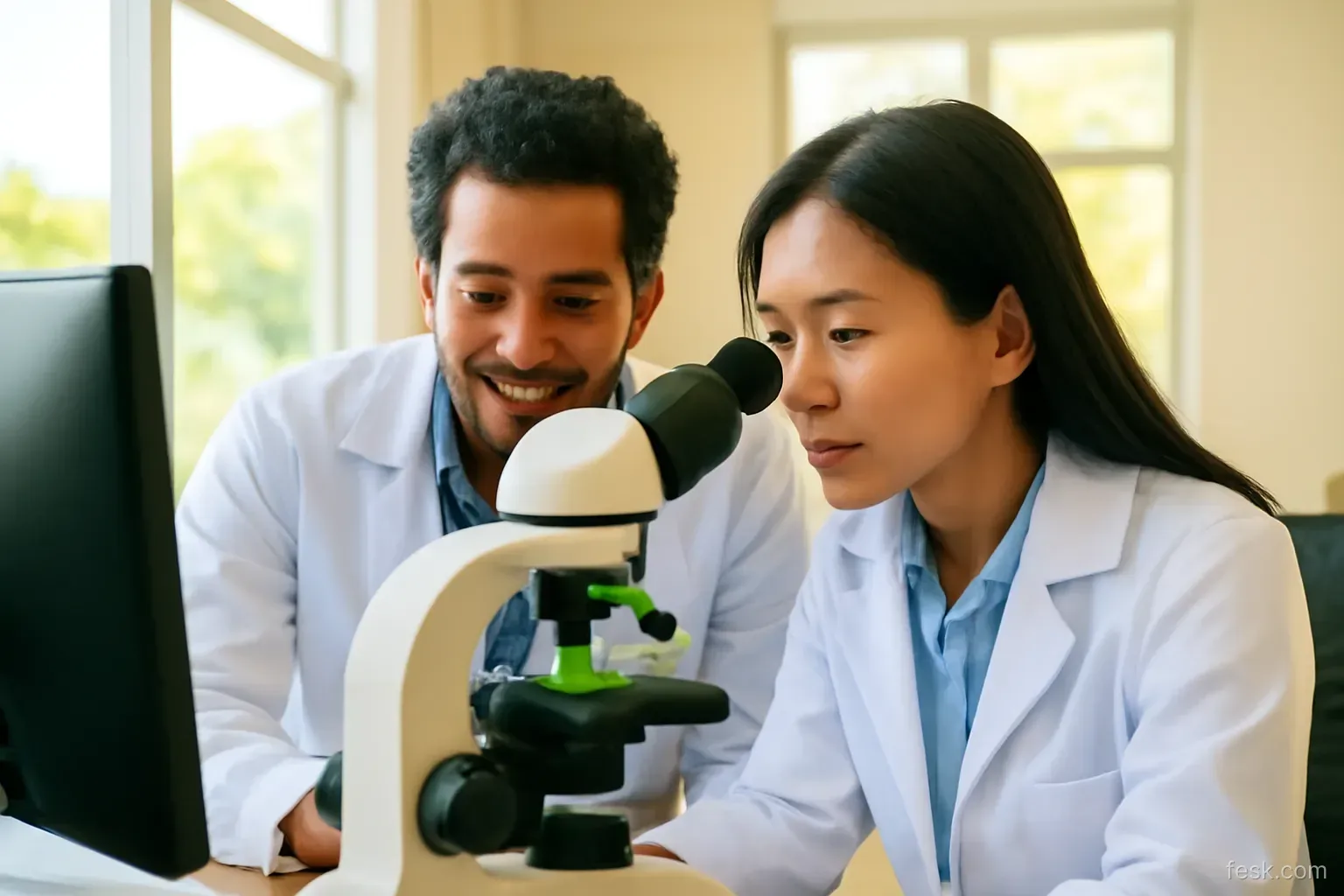
A molecular biology student photographs the same Western blot three times and every exposure has a blotchy background. He picks the cleanest, uses a clone tool to remove a smudge beside lane 4, then boosts contrast on the faint bands so reviewers can see them. Nothing was invented. Yet if an editor asks for the raw image, all three steps could be classed as inappropriate manipulation, and the paper could be rejected or corrected.
Images in a paper are data, not decoration. The core rule fits in one sentence: any edit that changes what a reader believes was actually recorded is off limits unless it is clearly declared. The rest of this article turns that sentence into concrete decisions.
Treating images as measurements
Once you see an image as a measurement, the question stops being “does it look good?” and becomes “does it still represent the original signal?” A faint band is information: it says the protein level is low. A background smear is information: it says something about the quality of the run. Removing either removes part of the result.
The same logic applies well beyond the wet lab: electron micrographs of materials, archaeological site photographs, satellite scenes, histology slides, even screenshots of interfaces in user research.
Adjustments that are usually acceptable
Journal guidelines and bodies such as COPE converge on a handful of principles:
- Apply changes to the whole image: brightness, contrast and colour balance across the full frame, not a selected region.
- Apply the same changes to controls: if the treatment image gets a contrast boost, the control image gets exactly the same one.
- Crop without hiding relevant information: trimming empty margins is fine; trimming to hide an unexpected band is not.
- Never make features vanish or appear: everything visible in the original remains visible, and nothing new is introduced.
A classification of common edits
| Edit | Status | Condition |
|---|---|---|
| Linear contrast change to the whole image | Acceptable | Applied equally to controls |
| Gamma or non-linear curve adjustment | Declare it | State it in the methods or figure legend |
| Cropping margins | Acceptable | Does not hide relevant bands or regions |
| Splicing lanes from different gels or positions | Declare it | Visible dividing lines and a legend note |
| Removing dust, smudges or bubbles | Not acceptable | Reimage instead |
| Cloning one region to cover another | Falsification | No exceptions |
| Reusing one image for two different conditions | Falsification | Even when rotated, flipped or recoloured |
| Enhancing contrast in one region only | Not acceptable | Unless declared and shown beside the original |
How to splice honestly
Splicing is not automatically wrong. You ran ten samples across two gels and want lane 3 next to lane 8 for comparison. That is fine, provided readers can see it is a composite:
- Insert a thin white or black line between the spliced pieces.
- State in the legend which lanes came from the same gel, which did not, and whether exposure times matched.
- Provide the full, uncropped gel images as supplementary material. Many journals now ask for these at submission.
If the two pieces come from different exposures, comparing intensities across them means little. Rather than splicing, rerun the samples you need to compare on a single gel.
Archiving raw images: the dull habit that saves you
When a question arises after publication, the first request from the editor is “please send the original images.” Researchers who can answer within a day tend to share these habits:
- Keep the raw file from the instrument, in its native format or uncompressed TIFF, never only an edited JPEG.
- Preserve metadata: acquisition date, exposure time, magnification.
- Name files with a convention that includes date, sample ID and experiment ID, and record that filename in the lab notebook.
- Edit only copies and record the steps. Tools such as ImageJ/Fiji can save a macro of every operation.
- Back up to at least two places, one of which is not your personal laptop.
How journals and readers check images
Many publishers now screen submissions with image-duplication software, and some employ dedicated research integrity editors for figures. After publication, images are also examined by sharp-eyed readers on post-publication commenting platforms. Typical signals include repeated background patterns, suspiciously crisp edges around a band, differing noise texture between halves of a panel, or identical cells turning up in two different figures.
Worth remembering: a large share of flagged cases are not deliberate fraud but wrong-file errors, made while assembling figures from a folder of hundreds of badly named images. File naming conventions and a notebook are your best defence.
Beyond the wet lab
- Field, heritage and specimen photographs: colour balancing is fine; removing people or objects, or replacing a sky, is not. Reconstructed images must be labelled as such.
- Materials micrographs: the scale bar must come from the original acquisition, not be redrawn by eye.
- Photographs of participants: blurring faces for anonymity is a legitimate edit and should be declared; you also need consent to use the image.
- Charts: never smooth a curve by hand in a graphics program; every transformation should come from the data and the code.
A 30-minute figure check before submission
- Build a table listing every figure, every panel, the matching raw filename and its acquisition date.
- Open each panel beside its raw file and compare. Any panel without a raw file gets a red flag.
- Check that no image appears twice in the paper or duplicates one in an earlier paper from your group.
- Reread the legends: splices noted, non-linear adjustments declared, scale bars present.
- Prepare a folder of uncropped originals ready to upload or send on request.
If you supervise students, make this check a mandatory step before any manuscript leaves the group. Ten minutes spent asking “where is the raw file for Figure 3B?” is far cheaper than a correction notice later.
Câu hỏi thường gặp
Is adjusting brightness on a Western blot considered misconduct?
No, as long as the change is linear, applied to the whole image and applied equally to the controls. Problems arise when one region is adjusted separately or when bands disappear or appear as a result.
Can I combine lanes from different gels in one figure?
Yes, provided the splice is marked with a clear dividing line and the legend states that the lanes come from different gels. Supplying the full uncropped gels as supplementary material is increasingly expected.
Is it okay to remove dust spots from a microscopy image?
Most journals do not accept local retouching, even of dust. The safe option is to reimage or leave the artefact and mention it in the legend if needed.
What format should I keep raw research images in?
Keep the instrument’s native format or uncompressed TIFF with its metadata intact. Do not rely on edited JPEGs, since each JPEG save discards information.
What should I do if I used the wrong image in a published paper?
Contact the editor promptly with the correct image, the raw file and an explanation of how the error happened. Self-reporting early usually leads to a correction rather than a harsher outcome.